cyto.site
Cytology, specimen prep, and the cell atlas
Welcome. I can help you find any cell, understand what you are seeing, or connect you with the right helper. What are we looking at?
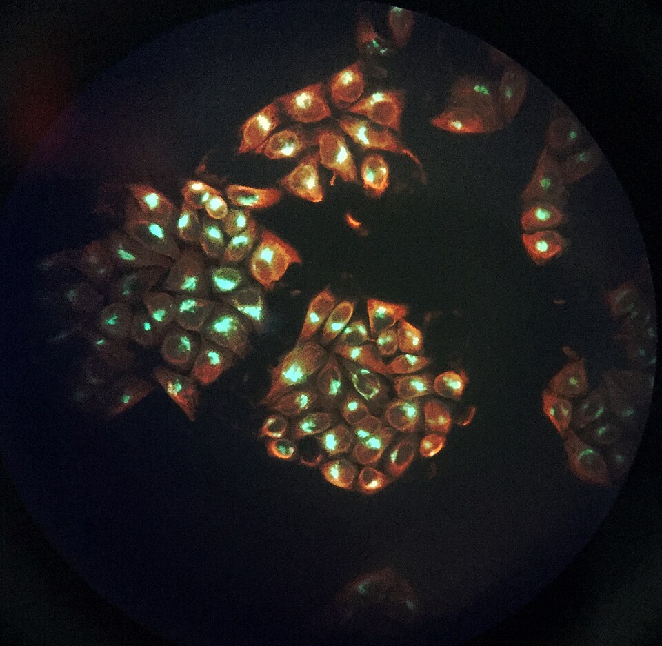
The cell on the front page is a human pluripotent stem cell, grown without feeder cells. Its lines were first derived from human blastocysts, and what makes it what it is are its markers, POU5F1, NANOG, SSEA-4 and TRA-1-60, and its clock: one division every 15 to 16 hours, with a G1 phase of only 2.5 to 3 hours, far shorter than a somatic cell's. It can form derivatives of all three embryonic germ layers, which is why it stands at the door of cyto.site. Sources: Thomson 1998, https://pubmed.ncbi.nlm.nih.gov/9804556/ ; Becker 2006, https://pubmed.ncbi.nlm.nih.gov/16972248/ ; Adewumi 2007, https://pubmed.ncbi.nlm.nih.gov/17572666/ ; Ponsaerts 2004, https://pubmed.ncbi.nlm.nih.gov/15671669/
Gallery 510
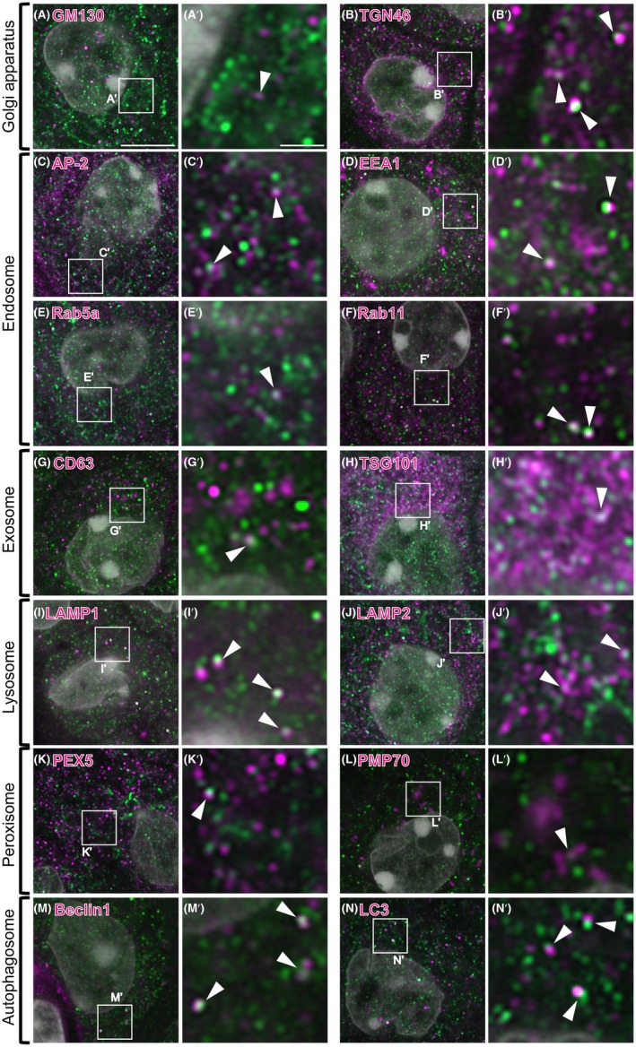
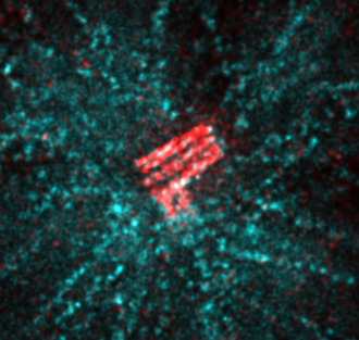
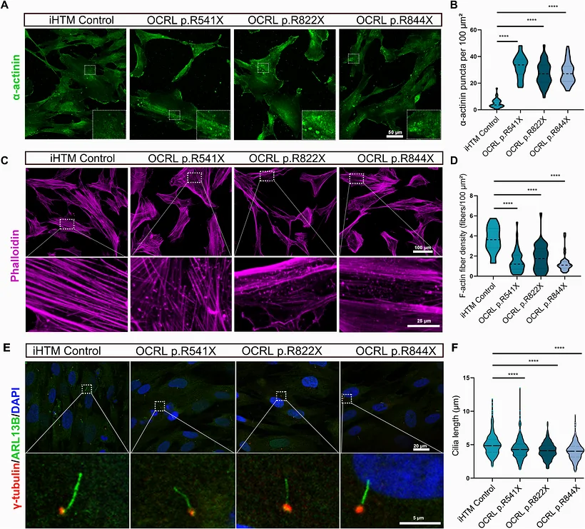
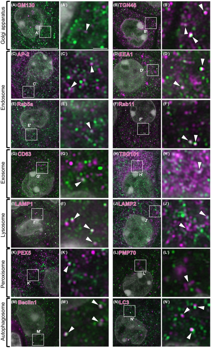
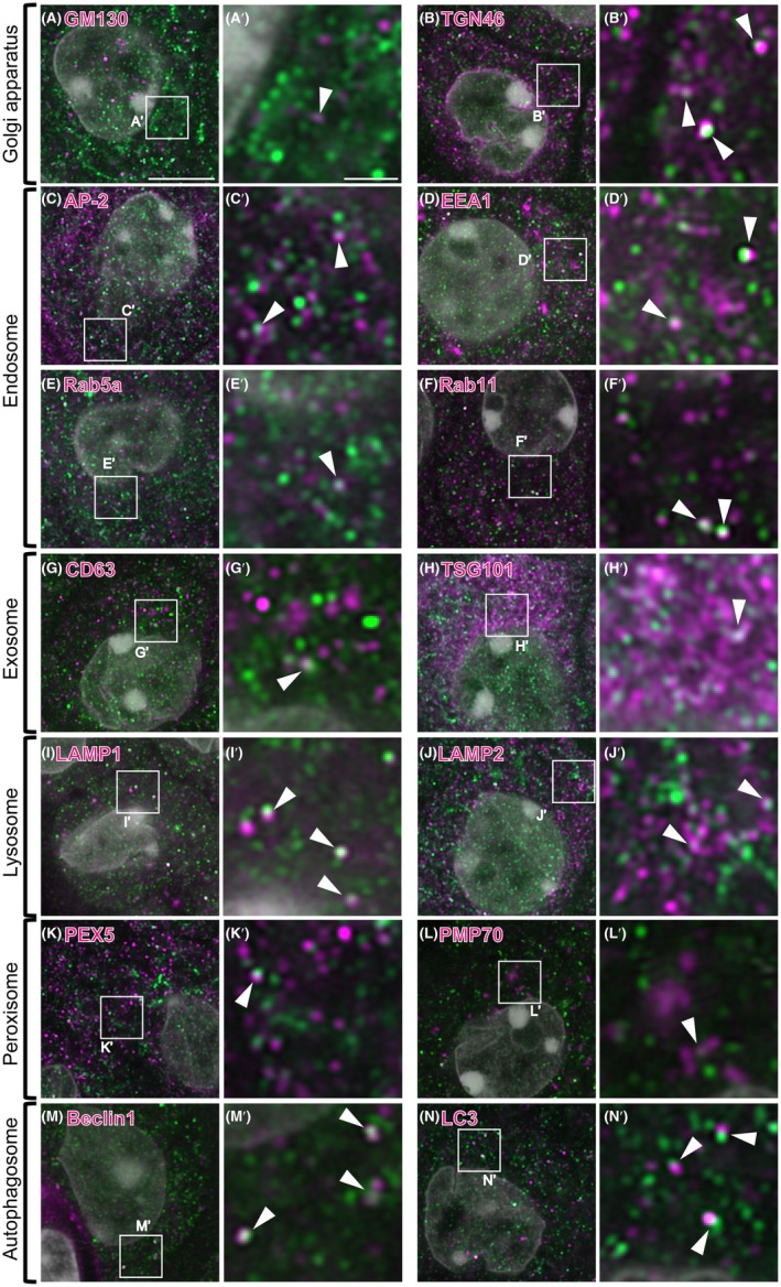
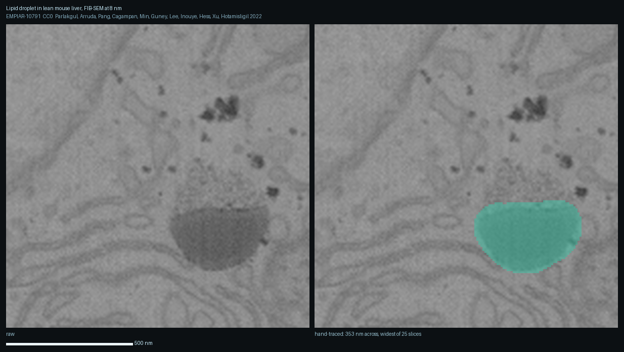
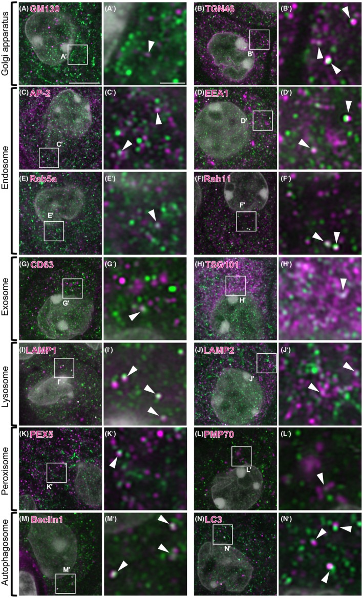
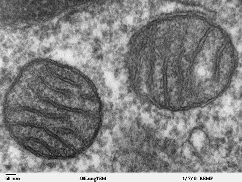
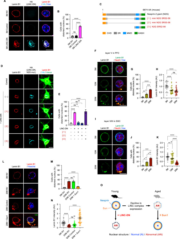
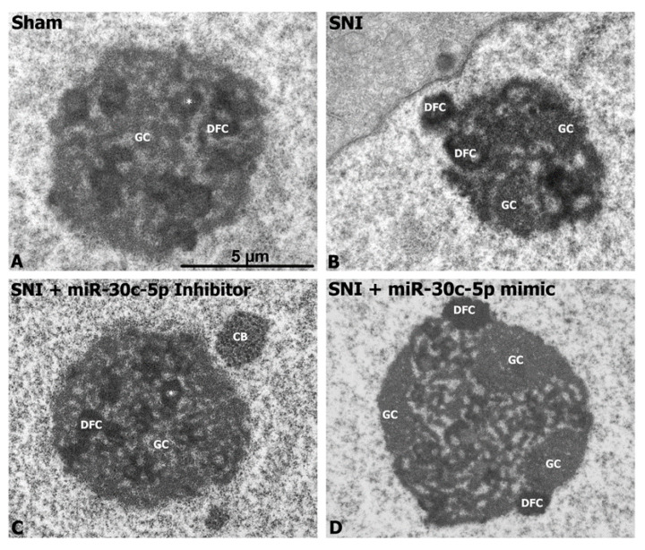
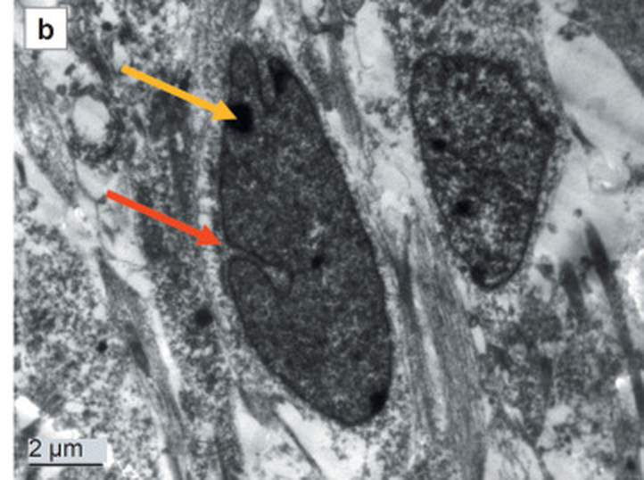
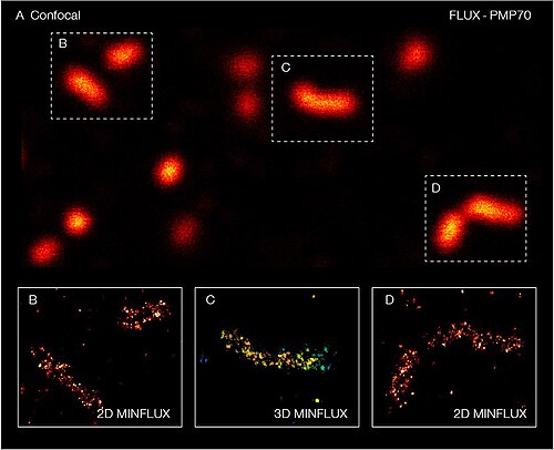
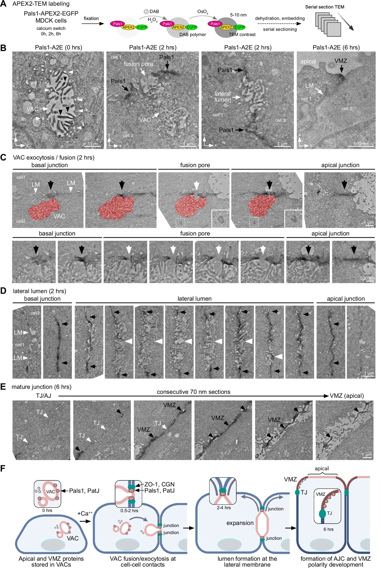
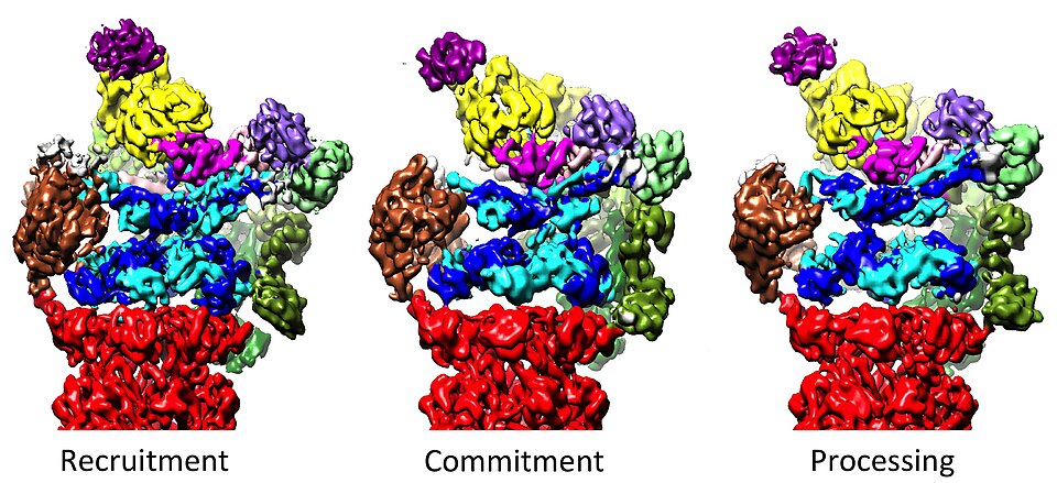
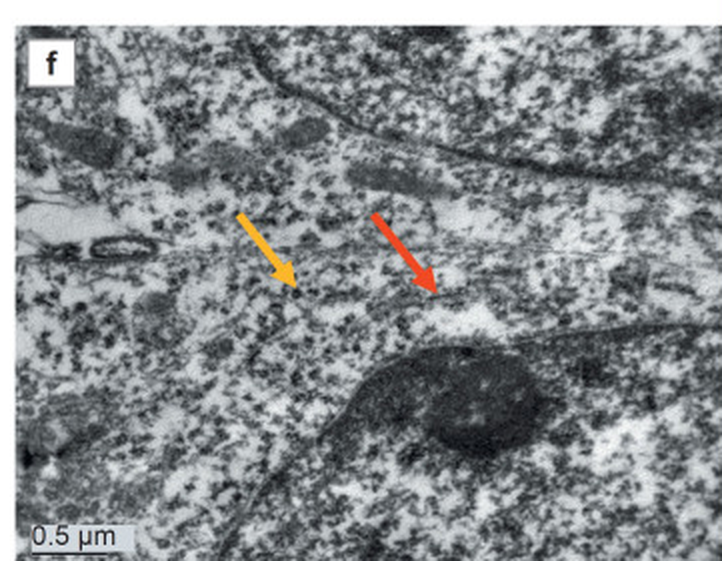
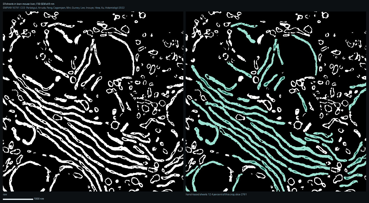
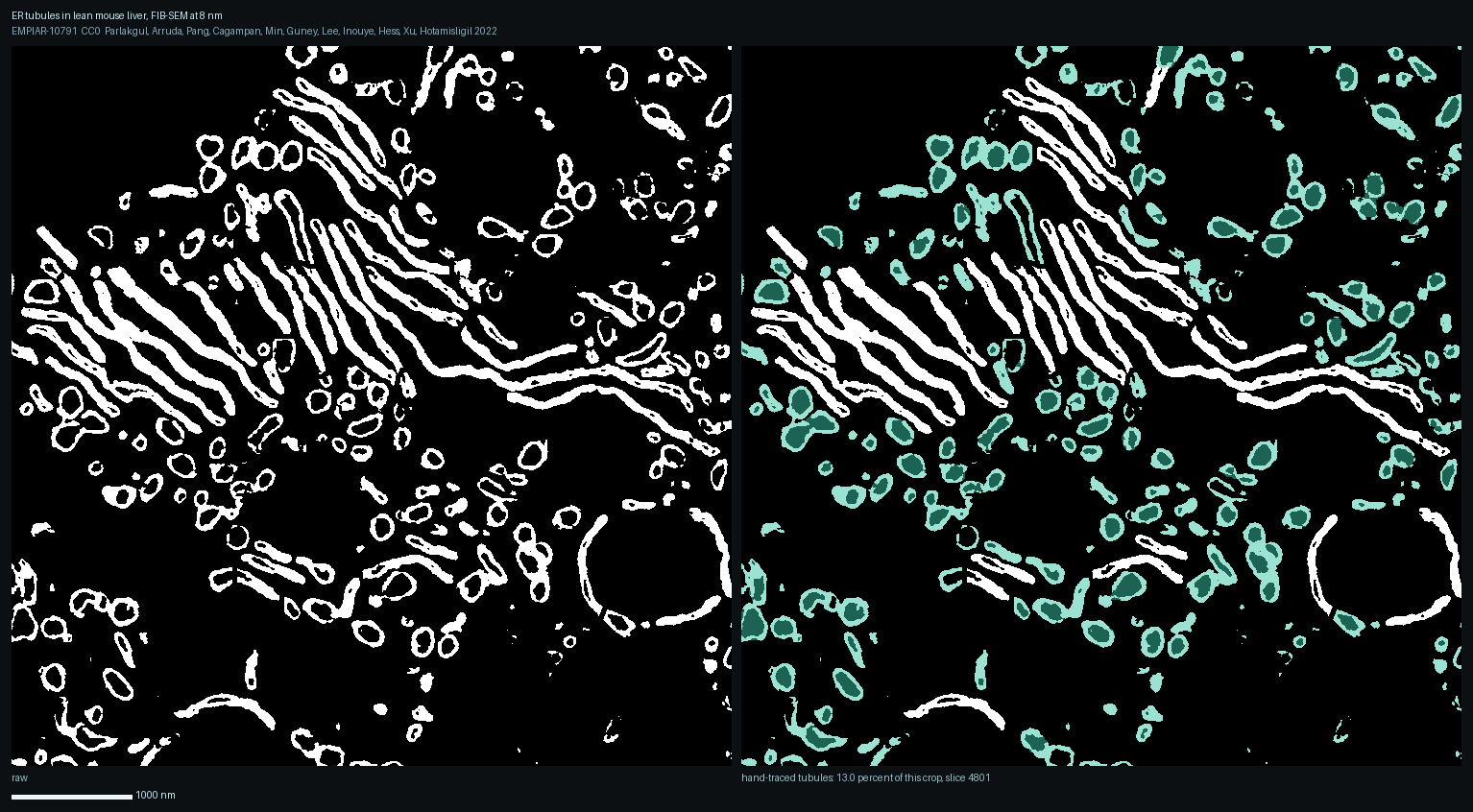
Full gallery →One picture per organelle here; the full gallery holds every picture from every cell site as well, filterable by cell and by organelle.
Library 8518
Open the library →Galleries and publication lists for every organelle and every cell: 8518 papers in Vancouver style, each PMID linked to PubMed, 23 cell galleries. Every PMID cyto.site cites is checked for retraction at each build; 2 retracted, each tagged.